Toxicity of Graphene Oxide Nanoparticles (Paper)
This study has me pondering how exposure to GO is absolutely unavoidable as this toxic substance is being promoted as the next most exciting thing – used in vaccines, drugs, air, food, clothes, agriculture, buildings, electronics, bandages, masks, and so on. My notes from the study and my initial-thoughts on what we might be able to do about it.
I was just going to reference this paper in my post on Graphene that I’m working on but I ended up taking so many notes that it needs it’s own page.
Rhazouani, Asmaa et al. “Synthesis and Toxicity of Graphene Oxide Nanoparticles: A Literature Review of In Vitro and In Vivo Studies.” BioMed research international vol. 2021 5518999. 10 Jun. 2021, doi:10.1155/2021/5518999 (01)
See also: Graphene toxicity as a double-edged sword of risks and exploitable opportunities: a critical analysis of the most recent trends and developments ( Yuri Volkov et al 2017 2D Mater) (02)
“Nanomaterials have been widely used in many fields in the last decades, including electronics, biomedicine, cosmetics, food processing, buildings, and aeronautics. They are currently used to administer drugs, proteins, genes, vaccines, polypeptides, and nucleic acids.
The natural routes of entry of nanoparticles into an organism are inhalation, ingestion, and dermal (skin exposure). Graphene oxide (GO), an oxidized derivative of graphene, is currently used in biotechnology and medicine for cancer treatment, drug delivery, and cellular imaging. (injection)
GO-based nanomaterials have been structured as topical antimicrobial media in the form of bandages, ointments, or cotton fabric. For this reason, dermal exposure is an important route of exposure that deserves attention.“
- GO is characterized by various physicochemical properties, including nanoscale size, high surface area, and electrical charge.
- The toxic effect of GO on living cells and organs is a limiting factor that limits its use in the medical field.
- There is no Standardization… “there is no specific method or procedure for producing a “standard” GO because each synthesis method produces a different GO type. Therefore, the GO has distinct physicochemical properties, such as structure and reactivity.”
- GO has a high adsorption capacity for proteins and antibodies.
- The toxicity of GO in cells is due to several factors, including dose, lateral size, and surface charge.
- To date, the studies carried out on the cytotoxicity of GO are contradictory. Some studies have shown that GO has no toxic effects on cellular behavior, while others have reported that this nanomaterial can induce cellular damage.
The toxic effects of GO on cells
Cytotoxicity of this nanomaterial in vitro is closely related to incubation conditions, including exposure dose, culture time, incubation temperature, and cell type. (So we can look at in vitro studies and evaluate it’s likely effects on us)
- Physicochemical properties of GO, such as shape, particle size, number of layers, and surface functionalization, affect the behavior of GO on cells.
- GO with a dose less than 20 μg/mL did not exhibit toxicity to human fibroblast cells, and the dose of more than 50 μg/mL exhibits cytotoxicity such as decreasing cell adhesion, inducing cell apoptosis, and entering into lysosomes, mitochondrion, endoplasm, and the cell nucleus.
- Yet, GO with a smaller size caused more severe oxidative stress and induced more obvious cytotoxicity in A549 cells compared to GO with a larger size.
- The interaction between the GO and the cell membrane can cause morphological changes and cell lysis, such as hemolysis of red blood cells (rupturing of red blood cells and the release of their contents into surrounding fluid). These changes are due to strong electrostatic interactions between negatively charged oxygen groups on the GO’s surface and positively charged phosphatidylcholine on the outer membrane of red blood cells.
- Incubation of human breast cancer cells MDA-MB-231 with GO caused a decrease in cell viability due to the dose of GO exposure.
- Toxicity of GO in the rat cardiomyoblast H9c2 cell line demonstrated that GO induced cardiotoxicity, mitochondrial disruption, generation of reactive oxygen species, and DNA interactions.
- Graphene oxide nanoparticles induced cytotoxicity in H9c2 cells.
- Graphene oxide nanoparticles induced mitochondrial hyperpolarization in H9c2 cells.
- Graphene oxide nanoparticles elicited an increase in free radicals in H9c2 cells.
- DNA damage was related to graphene oxide nanoparticles cytotoxicity.
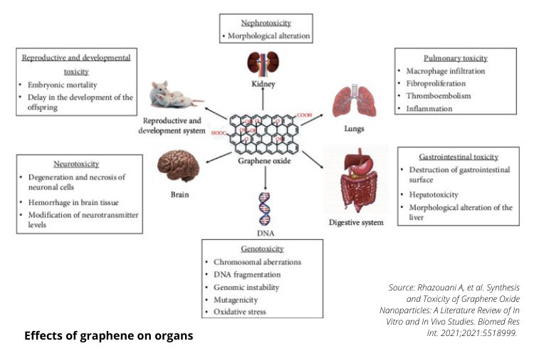
- Intratracheal (respiratory exposure) administration of GO in mice developed fibrosis in lung tissue after 21 days.
- GO increased the rate of mitochondrial respiration and the generation of reactive oxygen species, activating inflammatory and apoptotic pathways.
- Intravenous injection exposures could also be retained in the lung and induce the formation of granulomas and pulmonary edema.
- Inhaled GO nanosheets can destroy the ultrastructure and biophysical properties of pulmonary surfactant film, which is the host’s first line of defense, and reveal their potential toxicity.
- Once deposited at the bottom of the pulmonary alveoli, nanoparticles can be taken up by macrophages or eliminated by respiratory mucus via the action of hair cells or, for the smallest of them, pass through the pulmonary epithelium and end up in the interstitial liquid.
- Intraperitoneal (abdominal tissue/membrane) injection, the GO mainly remained accumulated near the injection site and small agglomerates (masses) could be found in the liver and spleen’s serous membrane. GO polyethylene glycol (GO-PEG) functionalized derivatives are mainly retained in the reticuloendothelial system, including the liver and spleen.
- GO particles are aggregated (massed) by interaction with digestive fluids and the acidic pH of the stomach.
- Low-dose GO caused severe damage to the gastrointestinal tract in maternal mice rather than high-dose GO. This is because the low dose of GO without agglomeration (process of collecting in a mass) can easily attach to the gastrointestinal surface and cause destruction by its abundant sharp edges.
- Bleeding in brain tissue was observed in animals with GO intoxication.
- GO is a nephrotoxic product and that its toxicity can be mediated by oxidative stress.
- Administration of GO significantly increased the activities of superoxide dismutase, catalase, and glutathione peroxidase in a dose-dependent manner in the kidney compared to the control group.
Neurotransmitters, DNA & Cell Membrane Damage
- Exposure to GO caused a significant decrease in neurotransmitters such as tyrosine, tryptophan, dopamine, tyramine, and GABA.
- Exposure to GO caused a decrease in the expression of ttx-1 and ceh-14 genes, which are genes necessary for sensory neurons’ functioning.
- Proposed that small nanosheets of GO reduce glutamate availability, which is the major excitatory neurotransmitter in the central nervous system.
- GO is capable of inducing genotoxicity. The use of GO at concentrations of 10 and 100 μg/mL altered gene expression.
- Injection of GO at different doses (10, 20, and 40 mg/kg) for one or five consecutive days, caused genomic instability, mutagenicity, and oxidative stress in the liver and brain tissue.
- Administration of GO significantly increased dose-dependent DNA breaks and induced mutations in the p53 (6 and 7) and presenilin (exon 5) genes by increasing the expression of the p53 protein.
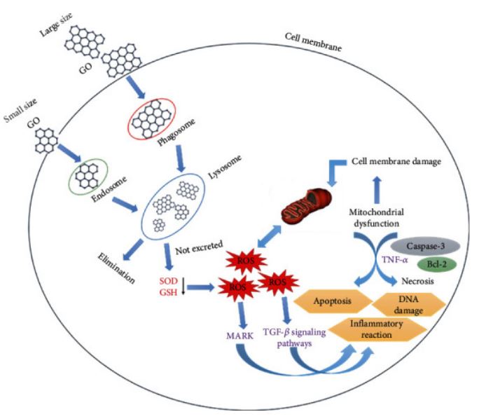
- Small GO could be degraded by lysosomes and eliminated from the body without causing observable toxicity.
- Large GOs could cause damage to the cell membrane by binding to proteins and interacting with phosphatidylcholine, leading to ROS production, and increasing the dose and duration of exposure to GO results in a progressive decrease in the activity of SOD and GSH.
(Does this image look strangely familiar to anyone else who studied SARS-COV-2 in the first year? or is it just me that sees the resemblance?)
Excretion
GO excretion varies in different organs. The routes of eliminating GO in vivo have not yet been clearly explained, but renal and fecal routes appear to be the major elimination routes. To date, several controversial results have been obtained regarding the distribution and excretion of this nanomaterial.
- Lungs = challenging to eliminate, causing inflammation, cell infiltration, granuloma formation, and pulmonary edema.
- Liver = can be eliminated through the hepatobiliary pathway by following the duodenum’s bile duct.
- GOPEG = GO polyethylene glycol’s functional derivatives accumulate primarily in the liver, and the spleen can be eliminated gradually, probably via the kidneys and fecal excretion.
- GO particles with large size of 200 nm are trapped by splenic physical filtration.
- Intratracheally (respiratory tract) instilled GO nanosheets can be retained in the lungs. This exposure resulted in acute lung injury and chronic pulmonary fibrosis. GO-induced acute lung lesions are related to oxidative stress.
- Intravenous (injected) administration of GO caused massive pulmonary thromboembolism in mice.
- Direct administration of GO into the lungs of mice resulted in severe and chronic lung damage. These GO nanosheets disrupted the alveolar-capillary barrier, allowing inflammatory cells to infiltrate the lungs and stimulate the release of proinflammatory cytokines.
- GO has a toxic effect on nerve tissue.
What this paper has me pondering…
This paper makes me really consider the urgency in needing to know exactly how (or if) it can be removed from the body, as it seems that it’s in everything and there is no avoiding it – it’s in vaccines, it’s in the food, the air, the water, clothing, drugs, masks, ointments, bandages, agriculture, and more.
It seems like this paper is pointing out the contradicting studies – and although they didn’t say it – I will – my guess is that the studies that show “favourable” results, most likely have pharma-funded backgrounds or a conflict of interest that needs the graphene studies to look good in order to get this cheap and toxic product approved for all kinds of things. This is how the pharmaceutical industry works, and the journal publications are their biggest marketing campaign and consumer-targets.
Looking at this paper in combination with the other studies I’ve read so far, it seems that the best we can do to mitigate this toxicity of this seemingly unavoidable substance:
- Avoid it as much as possible – that’s common sense – but if they end up putting it in our water, clothes, medicines, injections, and if they are putting it in the air and agriculture already – and promoting it to our governments as “the next best thing” – it may be something that none of us can avoid while greed and corruption rules the earth.
- Increase glutathione levels
- Eating high glutathione organic foods such as greens and high chlorophyll foods, broccoli, parsley, cabbage, garlic, brussels sprouts, beets, asparagus, avocado, cucumber, green beans, spinach, sunflower seeds, mushrooms, Brazilian nuts, tomatoes, and plant-based protein-rich foods.
- Supplementing with NAC & Glutathione
- Avoiding supermarkets and going straight to farm / growing our own foods without the nasty pesticides, sprays, packaging, and GMO’s – and their NWO-Foods.
- Eat a Mediterranean Diet as much as possible as that takes care of all the organs that may be effected, and also can be credited for pretty much all reversal of diseases and cellular-regeneration.
- Don’t eat anything in pretty packaging from large corporations. Don’t eat supermarket meat.
- Sulforaphane (Broccoli, Broccoli Sprouts, Cabbage, Cauliflower, Brussels Sprouts) to reset the genes and for its antioxidant, antimicrobial, and anti-inflammatory properties, cancer prevention, heart health, neurological issues and more. Add that to our diet daily.
- If these neurotransmitters are depleted (tyrosine, tryptophan, dopamine, tyramine, and GABA) – do we supplement them or find food-sources to compensate? My guess is that a Mediterranean diet with the addition of fermented foods (yogurt, kefir, etc.) will do the trick here, but I’ll look more into it.
- The lung-damage looks like one of the worst effects, but as someone who has repaired her lungs, I know this is not impossible to rectify (as long as you don’t listen to the in’doc‘trinated) as I have repaired my lungs from emphysema – everything is repairable if you don’t listen to genocidal maniacs who have control of the pharmaceutical, media, and agricultural industries (and more things make sense when you realize the planet is owned and governed and controlled by essentially one entity – even though it’s several hundred families – it’s still a division of the one entity that profits and enslaves us from our illnesses, woes, desires and fears).
- The vessel-damage all the way through the body could be the most damaging as it may be very difficult to detox/eliminate such tiny (nano) sized razor-sharp particles, so having a healthy diet may be our only saving grace to prolonging life and to have a faster cellular-regeneration process (as if it messes/roughs-up the cells as it slices it’s way through our linings, organs, vessels, etc., if we have a toxic diet, we’re going to be speeding up the possibility of problems, i.e. things getting stuck where it wouldn’t of previously got stuck as the lining was “smooth” before the GO).
Ponder on this some more I shall. This is just my initial thoughts after reading the study. Feel free to share your thoughts on Telegram.
See posts tagged: NWO-Food | Graphene
Related Studies to go through
Just want to keep some bookmarks here of relevant studies I haven’t had time to look through yet (so that I can close some open tabs lol).
- Differential genotoxic and epigenotoxic effects of graphene family nanomaterials (GFNs) in human bronchial epithelial cells (The effects of GFNs on cellular genome were evaluated with single and double stranded DNA damage and DNA repair gene expressions. Genotoxic and epigenotoxic potentialities of graphene family nanomaterials (GFNs). The GFNs possibly caused DNA damage by affecting NER and NHEJ repair systems. Possibly DNMT3B and MBD1 genes regulate GFN’s induced global DNA methylation status.) (03)
https://doi.org/10.1016/j.mrgentox.2016.01.006 - Graphene oxide nanosheets induce DNA damage and activate the base excision repair (BER) signaling pathway both in vitro and in vivo (Studies have reported that excessive GO exposure can cause cellular DNA damage through reactive oxygen species (ROS) generation. GO as a potential emerging pollutant is first investigated using atomic force microscopy (AFM). GO exposure is first confirmed to cause DNA damage when the concentration is higher than 25 μg/mL in HEK293T cells. BER pathway could be a possible inner response mechanism for GO exposure both in vitro and in vivo.) (04)
https://doi.org/10.1016/j.chemosphere.2017.06.049 - Short-term inhalation study of graphene oxide nanoplates (05)
- Toxicity of graphene-family nanoparticles: a general review of the origins and mechanisms (06)
- Toxicity of graphene and graphene oxide nanowalls against bacteria (07)
- Graphene Nanomaterials: Synthesis, Biocompatibility, and Cytotoxicity (08)
- Toxicity of graphene in normal human lung cells (BEAS-2B) (09)
- Can nanomaterials induce reproductive toxicity in male mammals? A historical and critical review (10)
- Graphene oxide affects in vitro fertilization outcome by interacting with sperm membrane in an animal model (11)
- Graphene Oxide increases mammalian spermatozoa fertilizing ability by extracting cholesterol from their membranes and promoting capacitation (12)
- Graphene and carbon nanotubes activate different cell surface receptors on macrophages before and after deactivation of endotoxins (13)
- Ex Vivo Human Colon Tissue Exposure to Pristine Graphene Activates Genes Involved in the Binding, Adhesion and Proliferation of Epithelial Cells (14)
- Safety Assessment of Graphene-Based Materials: Focus on Human Health and the Environment (15)
And if it’s “superparamagnetic iron oxide nanoparticles (SPIONs)“ that we’re dealing with:
- Potential toxicity of superparamagnetic iron oxide nanoparticles (SPION) (2010) (16) Cellular SPION overload may trigger adverse cellular responses and the long-term impact of these acute exposures are not well understood. Plausible that internalised SPION may corrode over a long period of time by releasing metallic ions that in turn bear a long-established correlation with DNA damage. Ideally, it would be worthwhile to decipher the stability and breakdown products of coatings because a ‘biocompatible coating’ that is considered stable initially may eventually break down into an unfavourable product or expose the iron oxide core, thereby eliciting adverse cellular responses. Emerging studies have begun to highlight aberrant cellular responses including DNA damage, oxidative stress, mitochondrial membrane dysfunction and changes in gene expression as a result of SPION exposure, all in the absence of cytotoxicity.
DOI: 10.3402/nano.v1i0.5358 - Superparamagnetic iron oxide nanoparticles: magnetic nanoplatforms as drug carriers (2012) This 2012 study published in the International Journal of Nanomedicine talks about a “super-magnetic” phenomenon used in biomedical fields that once the drug is loaded is “guided by an external magnet” to their target tissue. (17)
- Magnetic Nanoparticles in Human Health and Medicine: Current Medical Applications and Alternative Therapy of Cancer (Book) (2021) “… valuable resource for researchers in the fields of nanomagnetism, nanomaterials, magnetic nanoparticles, nanoengineering, biopharmaceuticals nanobiotechnologies, nanomedicine,and biopharmaceuticals, particularly those focused on cancer diagnosis and therapeutics.” (18)
Other Related Tabs I want to close for now but look at later:
- Graphene Oxide and Covid Vaccines By Craig Paardekooper (PDF) (19)
- Toward the Application of High Frequency Electromagnetic Wave Absorption by Carbon Nanostructures (20)
- Nanotoxicology: Breathing carbon nanotubes causes pulmonary fibrosis, a cause of lung cancer (21)

Site Notifications/Chat:
- Telegram Post Updates @JourneyToABetterLife (channel)
- Telegram Chatroom @JourneyBetterLifeCHAT (say hi / share info)
- Gettr Post Updates @chesaus (like fakebook)
Videos:
References


